Which journal is the 5th most-cited worldwide, with over 734,000 citations in 2023? And which offers authors a prestigious place to publish their research, belongs to the Nature family, and provides open access?
It’s the same title – Scientific Reports – where Artur Dobrowolski, our young researcher from SiC Technologies Research Group, published his article. Together with Jakub Jagiełło, Beata Pyrzanowska, Karolina Piętak-Jurczak, Ewelina B. Możdżyńska, and Tymoteusz Ciuk, they prove once again that innovative methods in materials imaging are the future!
Our scientists described in the article PRISM: three-dimensional sub-diffractive phase-resolved imaging spectroscopic method | Scientific Reports (nature.com), the innovative PRISM method for material imaging developed by their team. This method allows for the creation of three-dimensional images of various types of samples – both two-dimensional and three-dimensional, as well as hybrid samples – and enables the analysis of material structures with unprecedented precision.
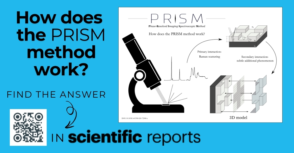
The PRISM method works by analyzing selected Raman-active modes, which allows for obtaining information about the material phases of the sample. In this method, specific light signals characteristic of different materials are detected. The data is collected in the so-called back-scattering geometry – meaning that the light hitting the sample is reflected back toward the detector. The entire measurement is performed using light with a specific wavelength – 532 nm – which is typical for green lasers. The protocol, or the titular PRISM (Phase-Resolved Imaging Spectroscopic Method), allows for sub-diffractive and material-resolved imaging of the object’s constituent material phases. The novelty of this method lies in the fact that spatial imaging can be achieved in two ways:
- Direct Mode uses the signal distal attenuation ratio, which enables the acquisition of spatial images.
- Indirect Mode relies on more subtle light-matter interactions, such as interference enhancement and light absorption, allowing for detailed analysis of the internal properties of the material.
The phase component is brought about by scrutinizing only selected Raman-active modes. We illustrate the PRISM approach in common real-life examples, including photolithographically structured amorphous Al₂O₃, reactive-ion-etched homoepitaxial SiC, and Chemical Vapor Deposition graphene transferred from copper foil onto a Si substrate and AlGaN microcolumns. The method is implementable in widespread Raman apparatus and offers a leap in the quality of materials imaging. The lateral resolution of PRISM is stage-limited by step motors to 100 nm. At the same time, the vertical accuracy is estimated at a nanometer scale due to the sensitivity of one of the applied phenomena (interference enhancement) to the physical property of the material (layer thickness).


